Written by: Ashley Wagner, Ph.D., Director of US Business & Technical Sales Probiotech International Inc.
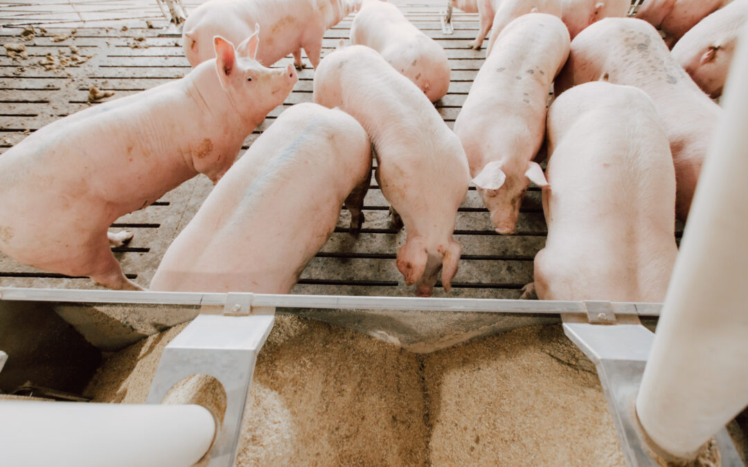
Weaning is a highly stressful event for pigs.
Intestinal health is crucial for pig well-being and performance.
CCC, a blend of monoterpenes and polyphenols, can mitigate weaning stress.
Improved intestinal health through CCC leads to better pig performance.
Trial Report: Impact of CCC on impaired intestinal integrity due to weaning
Pigs experience environmental, physiological, and social challenges even under the best management practices in today’s pig production systems. These challenges elicit a stress response in the brain that signals biological systems away from homeostasis. The severity of the challenge will result in differing impacts to the animal. For example, a social challenge may be mixed in open pen gestation and could lead to aggressive behaviors that may have no economic bearing or could be severe enough to lead to mortality. Although there are several challenges in today’s pig production systems, weaning is by far the greatest challenge and most stressful period for the animal. This is because weaning encompasses many stressors into a single event.
Beyond the role of digestion and absorption, the intestines operate as a defense mechanism against pathogenic bacteria and toxins. The intestinal barrier functions through physical, chemical, and immune protective mechanisms. The physical barrier comprises epithelial cells and the tight junction protein complexes (Figure 2) that selectively allow the absorption of nutrients while preventing access to pathogens and toxins. The chemical barrier prevents the colonization of pathogens via antimicrobial proteins and mucins secreted by the epithelial and goblet cells, respectively. The intestinal immune barrier protects the animal by recognizing foreign stimuli at a local level. Weaning impairs intestinal integrity, compromising the intestines’ ability to perform these barrier functions.
Alterations to the physical barrier following weaning have been well-examined in pigs. Researchers have different tools to determine the integrity of the physical barrier of the intestines. The use of the Ussing chamber system has allowed researchers to evaluate intestinal permeability by measuring transepithelial electrical resistance (TER; Smith et al., 2010). This system takes living intestinal tissue harvested from pigs, allowing researchers to examine transport barrier function and leaky gut in real time. Leaky gut occurs when there is an impairment to the intestinal physical barrier (Figure 3). Another tool researchers have is measuring the protein or mRNA expression level of the tight junction protein complexes from intestinal tissue samples. A lower expression or level of protein indicates a leaky gut. Weaning stress alters intestinal TER (Boudry et al., 2004; Smith et al., 2010; Hu et al., 2013) and decreases the expression of the following tight junction proteins: ZO-1, occludin, claudin-1, and -3 (Boudry et al., 2004; Hu et al., 2013; Cao et al., 2018).
Weaning as an event causes the pig to experience environmental, physiological, and social challenges. This alters the production of neurotransmitters utilized to signal from the brain to other biological systems that there is a stress event. There is a reduction in the amount of serotonin (a neurotransmitter involved in sleep, hunger, and general good feelings), and an increase in norepinephrine and epinephrine which work together to mobilize the body for fight or flight. This alters normal biological systems from homeostasis. Additionally, there is a rise in the stress hormone cortisol, which aids in altering normal body functions. These neurotransmitters signal many other pathways that there is a challenge (weaning), which eventually leads to alterations in digestion, absorption, and the barrier function of the intestines previously described.
So, how do we overcome what the body naturally does to set these pigs up for a successful transition during weaning?
CCC
CCC (Essential Ag Solutions, Sioux Falls, SD) is a blend of monoterpenes and polyphenols expertly selected for their anti-anxiety, anti-depressant, and antioxidant properties. Monoterpenes and polyphenols are compounds found in many plants, such as herbs (oregano, eucalyptus) and fruits (citrus and grapes). Monoterpenes are volatile compounds made up of terpene isomers. These compounds’ volatility produces an aroma that alters brain signaling. This effect is amplified when animals both smell and consume monoterpenes. This blend of compounds in CCC increases serotonin production (Perveen et al., 2009) while decreasing circulating cortisol production during a stress event such as open pen gestation (Brown et al., 2017). Together, this lowers the systemic stress response on other biological systems.
We recently completed a trial to evaluate CCC’s impacts on pigs’ intestinal integrity following weaning. This trial utilized 1296 weaned pigs from the same sow farm. Pigs were fasted during a 1-hour transportation to a commercial wean to finish the research facility. Pigs were assigned to either a control or CCC treatment: 24 pens per treatment and 27 pens per pen. Nine pigs per treatment were sacrificed on day 7 for ileum sample collection to determine tight junction gene expression. Initial body weights did not differ between treatments, but d 7 body weights were heavier in the CCC group. There was also an improvement in body weight gain during the first seven days for CCC pigs over the control. The day 7 gene expression for the tight junction proteins ZO-1, Claudin-1, -2, and -4 was elevated in the CCC group compared to the control by 49%, 151%, 232%, and 192%, respectively. The combined results of improved intestinal integrity and higher body weight at the end of 7 days indicate the ability of CCC to assist the weaned pig in a successful weaning transition.
Weaning stress is a challenge that all pigs endure. As a producer, the goal is to get them thriving quickly. This will enhance profits as well as the animal’s performance. By reducing the perception of weaning stress with CCC, intestinal integrity is less impaired. This may reduce the susceptibility to additional challenges (microbial) that may further impair performance. CCC is the key to getting over the weaning slump. Ask your Essential Ag Solutions representative about CCC; it is available in both liquid and feed form.
Written by: Manuel Contreras, DVM, MS, Diplomate ACPV, Agrimprove/Special Nutrients
KEY TAKEAWAYS:
Endotoxins are only present in Gram-negative bacteria.
Endotoxins can affect commercial poultry when some bacterins are injected without following the manufacturer’s instructions.
There are not many scientific reports currently reporting impaired performance, lesions, and symptoms associated with the presence of endotoxins in Poultry.
The importance of endotoxins in poultry production is a new topic frequently covered in animal production and veterinary medicine technical magazines worldwide. Marketing campaigns are, in part, responsible for this trend. Some commercial/technical articles show the effect of several mycotoxin binders against endotoxins. Reviewing some of these reports, we quickly detected the absence of measurements indicating that endotoxins were present in the evaluated animals. Therefore, it is worth asking how we can associate performance improvements when using these additives if, during the trials, the presence or reduction of endotoxins was not reported. We fully understand that a significant limitation in detecting endotoxins is the lack of availability of reliable laboratory tests in blood, urine or feces. So far, the techniques available are inaccurate, and the interpretation of the results tends to confuse researchers. In the last decade, we have evaluated the results of field tests carried out in pig farms in the European Union, where endotoxins were suspected of causing production deficiencies. In this case, a mycotoxin binder was evaluated to ameliorate the poor performance reported.
Although a statistically significant effect was reported in various production parameters when using the additive, it was not clearly established that its inclusion in the feed caused the changes.
What are endotoxins?
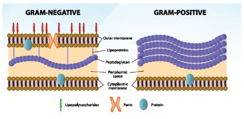
Endotoxins are used to describe a complex containing lipopolysaccharides (LPS) associated with the outer membrane of bacteria such as Escherichia coli, Salmonella, Pseudomonas, and Pasteurella.
Gram Negative bacteria have a cell envelope that contains three layers or membranes:
Cytoplasmic membrane (interior)
Peptidoglycan membrane or R layer
Outer membrane
Since Gram-positive bacteria do not have an outer membrane, endotoxins are not present.
How are endotoxins released?
1. Production during the initial bacterial growth phase, both in the laboratory (in vitro) and in animals (in vivo).
Through this mechanism, minimal amounts of soluble endotoxins are released. It is important to emphasize that for this growth and release to occur, a liquid medium is needed. Once released from the cell wall, an immune response is initiated that depends on the type and concentration of LPS, duration of exposure, host genetics, and the presence of clinical signs caused by viral or bacterial infections.
2. Destruction (lysis) of bacteria by the immune system or antimicrobial agents.
Since the intestines are loaded with Gram-negative bacteria, they represent animals’ most significant source of endotoxins. Its elimination in the feces allows it to combine with food and form a bioaerosol that can stimulate an inflammatory response in the respiratory tract. In experimental tests, when performing intra-tracheal inoculation of endotoxins in broilers, hypertension of the lungs was reported, and it is speculated that it may play an essential role in the development of ascites.
To emphasize the excellent dissemination capacity of this bio-aerosol in commercial poultry production, the same type of endotoxin present in birds has been detected in the blood of farm personnel.
The most important bacteria containing endotoxins belong to the group Enterobacteriaceae, which inhabits the normal intestinal microflora of birds and mammals (including humans).
Composition of LPS:
Lipid A
Nucleus
O antigen
Lipid A is a hydrophobic structure not mixed with water and is associated with toxicity. It acts as an anchor when bacteria invade the host’s cells. Even though endotoxins differ, Lipid A is always the same regardless of the bacteria. This factor explains why endotoxins from different bacteria cause the same type of damage in the host.
When animals are exposed to a stressful environment, the concentration of free endotoxins in the body increases. Once the balance established in the intestinal microflora is lost (dysbacteriosis or dysbiosis), the result is the development of Salmonellosis and/or Colibacillosis. In other words, the absence of some beneficial bacteria, such as Lactobacillus, will allow pathogenic bacteria to grow in the intestines.
It is essential to differentiate endotoxins from exotoxins. Gram-negative and Gram-positive bacteria produce the latter and mainly affect the host. Unlike endotoxins, exotoxins are secreted by bacteria in small amounts and are lethal.
Can endotoxins cause harm to commercial birds?
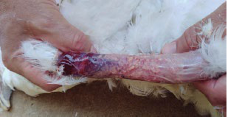
Deleterious effects have been observed when injecting bacterins prepared with Gram-negative bacteria for decades. Scientific publications have shown that by injecting small concentrations of endotoxins from Pasteurella multocida in broilers, the clinical signs of acute cholera can be reproduced without the need to challenge using the whole bacteria. The best example of this type of damage at a commercial level is caused by the injection of bacterins against Fowl Cholera, which depressed birds characterize after its application in pullets during the rearing period.
On the other hand, the reactions at the application site are significant due to condemnations at the slaughterhouses, especially when using oil emulsions. A recommendation by biological companies that illustrates the importance of endotoxins when vaccinating is to keep vaccines at an average temperature of approximately 37 ºC. Overheating will release the endotoxins present in the vaccine, and mortality and hemorrhagic syndrome could occur.
Photo 1. Local reaction at the application site of a bacterin injected into the neck of a 12-week-old commercial pullet, probably caused by endotoxins (LPS). The photo was taken at 18 weeks and shows yellowish caseous material of bacterial origin due to needle or syringe contamination.
How are endotoxins prevented? Determining if they are harming birds before investing in prevention is critical. The following strategies can be used if a real deleterious effect is established.
Vaccination with endotoxin segments. Lipid A has been used experimentally to obtain protection. The big drawback is the high cost of these products.
Use of mycotoxin binders mixed with the feed (organoclays). In in–vitro tests, about 90% adsorption capacity against endotoxins is reported. However, this does not mean they necessarily work when used in animal feed. Several scientific tests conducted with pigs in the United States, challenged with pathogenic strains of E. coli, have demonstrated the efficacy of some clays in reducing the incidence of diarrhea and poor performance. Although endotoxins are not measured in these reports, it is speculated that part of the efficacy of these products is a consequence of neutralizing the endotoxins released by the bacteria used to challenge.
It is critical to follow the manufacturer’s recommendations when applying bacterins to prevent the impact of endotoxins.
In conclusion, although the adverse effects caused by endotoxins in pigs and dairy cows seem to be more established, their negative impact on poultry is not categorically demonstrated.
Written by: Ashley Wagner, Ph.D.1, and Olivia A. Martin, MBA, PAS2 written exclusively for Essential Ag Solutions
KEY TAKEAWAYS:
Readers will learn that ASFV has several mechanisms of transmission and that enhancing biosecurity measures is a necessary component of the protection toolbox.
Feed is one of the greatest on farm risks to contaminate a farm with ASFV.
Viral mitigants as a component of the diet are meant to reduce the viability of the virus before it gets to the farm.
Research on Virabloc has demonstrated a reduction in virus viability of the following viruses: PEDV, PRRSV, EhV, and ASFV.
In part one of this series, readers were introduced to two mega viruses, the African Swine Fever Virus (ASFV) and Emiliana Huxleyi virus (EhV). These mega viruses are very similar to each other in size, structure, and the way they infect the host cell, but EhV presents zero threat to humans or pigs, which makes it a safe surrogate virus for laboratory experiments in Dr. Declan Schroeder’s virology laboratory at the University of Minnesota. Declan C. Schroeder, Ph.D. is an Associate Professor at the University of Minnesota in Molecular Virology and Veterinary Population Medicine. His lab studies the viability and infectivity of many viruses. In our previous article, we discussed with Dr. Schroeder how temperature and holding times do little to inactivate either of these megaviruses. If you missed PART ONE, you can check it out here.
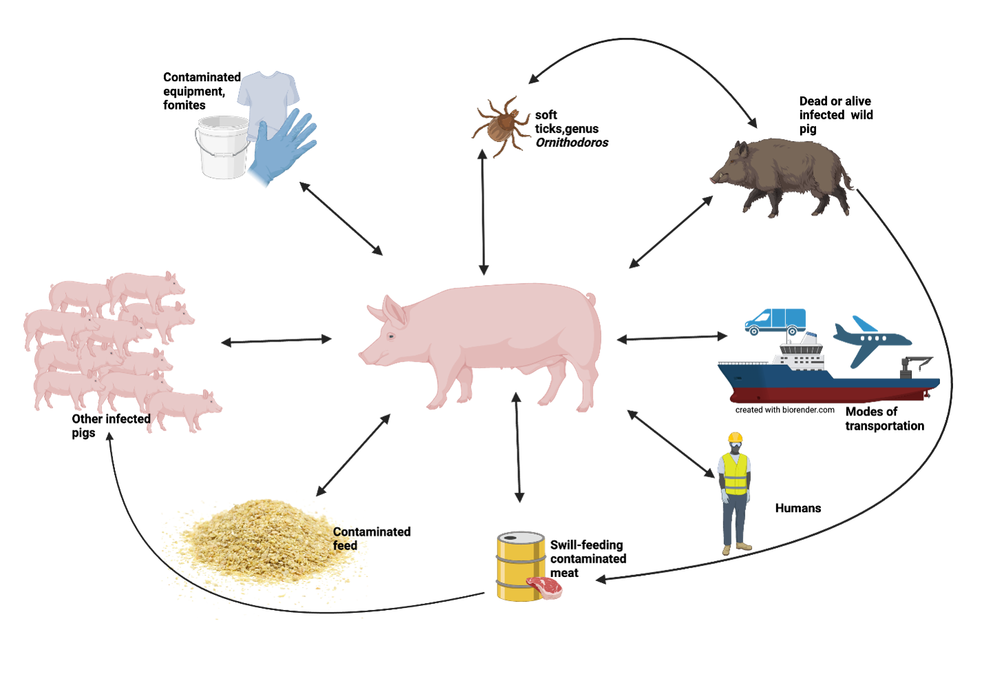
As of February 2024 (when this article was written), ASFV has not yet been detected in North America but has been detected in some Caribbean countries. With the threat of ASFV that close to the US, understanding how the virus spreads is key to protecting our swine industry. ASFV can spread through several mechanisms, some of which can be dealt with through enhanced biosecurity (see Figure 1). For example, contaminated equipment or even clothing could present a risk; movement of infected pigs or meat products from infected pigs could also play a role in spreading the disease. This is where both government control and individual farm biosecurity come together to protect the US swine industry. But the hidden transboundary threat that is introduced onto all farms is feed. Many feed ingredients are produced or sourced from countries that are ASFV positive, and as mentioned in PART ONE, ASFV can survive for long periods of time on feed ingredients. Therefore, feed could be both our greatest threat of infection and our greatest asset for protection.
There have been documented cases of infection in the US with lesser robust viruses, like PEDV and Seneca Virus A, where contaminated feed has been cited as the vector of transmission. Keeping in mind from Part 1 of this article series, Dr. Schroeder mentioned that his laboratory has demonstrated that ASFV survives during extreme temperatures that would destroy PEDV and PRRSV. Therefore, it is scientifically plausible that feed could be the method of entry for ASFV into the US swine industry.
In our early work with Dr. Schroeder, we investigated various feed ingredients and feed ingredient combinations and their effect on the viability of the surrogate virus for ASFV, EhV. Some of these ingredients included botanical extracts, spices, and medium-chain fatty acids. This initial work led us to the most active combination of ingredients for reducing EhV viability, commercially referred to as Virabloc (Essential Ag Solutions, Sioux Falls, SD, USA). Once Virabloc was developed, we continued our work to determine the best inclusion rate. In these experiments, 10, 5, and 2 lb per ton were tested against the viability of EhV. It was determined that Virabloc, at the lowest inclusion rate, significantly reduced EhV particle viability!
Virabloc has excellent success in the laboratory. However, our work with Dr. Schroeder aims to produce a combination of ingredients that can inactivate the virus in the feed so that it poses less of a threat on the farm. We have investigated the impact of Virabloc on reducing the viability of ASFV, EhV, PEDV, and PRRSV in animal feed. We have also looked at the impact of Virabloc over time. These trials demonstrated that Virabloc mixed with feed contaminated with ASFV, PEDV, and PRRSV (note: each trial was conducted on a single virus, not a combination) reduced the viability of the virus by 2 logs (99% reduction) after 1 hour of incubation. Additionally, the time course work demonstrated further reduction of both ASFV and EhV viability with time. These tests examined particle viability following 5, 10, and 21 days. The inclusion rate in all of these trials was 2 lb/ton, which is the manufacturer’s recommendation. How might this translate into a real-world situation? This means that if a feed ingredient in the feed mill is carrying ASFV particles, and Virabloc is added to the diet at the mill, Virabloc will be able to break down the virus before it gets to the farm!
It is important to remember that Virabloc is not a silver bullet but a tool in your protection toolbox that also contains good management practices and enhanced biosecurity measures. Although there is still more to uncover about the infection capacity of ASFV, researchers have determined that feed contaminated with the virus can serve as a potential route of introduction and transmission of ASFV into the United States, and that would be devastating to the swine industry. Therefore, virus mitigation strategies in the feed may be our greatest opportunity for protection against ASFV.
Contact your Essential Ag representative for more information on Virabloc or any other Essential Ag product to enhance production on your farm.
Figure 1. African Swine Fever Virus transmission vectors that could impact the US swine industry.
1Director of US Business & Technical Sales, Probiotech International 2 Marketing Director & Technical Sales Specialist, Probiotech International
Written by: Josep Garcia-Sirera, Special Nutrients Agrimprove
KEY TAKEAWAYS:
Endotoxins have a wide variety of effects on livestock and negatively affect production performance.
Different methods of endotoxin mitigation have been tested, but most are too expensive to be considered in animal production.
A more cost-effective method would be using toxin binders to capture endotoxins in the gastrointestinal tract and prevent them from entering the blood systems.
What are Endotoxins?
Gram-positive and Gram-negative bacteria are differentiated, among other things, by their type of cell envelope. Gram-negative bacteria are encircled by a thin peptidoglycan cell wall that is also enclosed by an outer membrane containing lipopolysaccharides (LPS), also called endotoxins. Gram-positive bacteria do not have an outer membrane; therefore, endotoxins are absent in this group. Gram-negative bacteria have a cell envelope that contains three essential layers or membranes: cytoplasmic (inner), peptidoglycan or R-layer, and the outer membrane. The latter contains phospholipids, proteins, and LPS. On the other hand, LPS consists of three elements: Lipid A, a hydrophobic component that serves as an anchor when a bacterium invades a host’s cell. The core is an oligosaccharide, and the O antigen is a hydrophilic component. Lipid A is apparently responsible for most of the toxicity caused by endotoxins.
Figure 1. Differences between Gram-negative and Gram-positive bacteria
Figure 2. Structure of LPS or endotoxin
Endotoxins are common in the gastrointestinal tract (GIT) of animals, particularly in the large intestine. They are also present in bioaerosols that produce inflammatory reactions, particularly in the respiratory tract. It has been detected that bioaerosols, coming from feed, litter, and feces, have the highest concentration of bacterial endotoxins. When endotoxins are present in the GIT and excreted together with feces, these endotoxins will be attached to the dust particles present in the farm and end up in the air, which causes the inhalation of particles by the animals. As a result, we can observe respiratory or gastrointestinal diseases affecting the animals that inhale them.
Under normal commercial production conditions, a small number of endotoxins will be transferred from the GIT to the bloodstream. According to scientific papers, it represents concentrations lower than ten pg/ml (picograms per milliliter), and these levels are capable of stimulating the immune system. Under stress, the level of free endotoxin in the GIT will be higher. Stress can produce dysbiosis or dysbacteriosis, that is, microbial imbalance in the intestines and the production of more endotoxins in the system. It is critical to emphasize the importance of keeping the correct microbial balance in the intestines because diseases such as Salmonellosis or Colibacillosis are intrinsically associated with the unrestricted growth of pathogenic bacteria that displace the intestinal favorable microflora (i.e., Lactobacillus to mention).
When bacteria are eliminated due to the administration of antimicrobials or because of the work of the immune system, bacterial cells will be destroyed, and the final consequence is the liberation of endotoxins that will harm the animal. Endotoxins are released from the bacterial cell wall during the growth and division phases of the microbe.
Understanding the Mode of Action
The mechanism is complex. In humans, LPS binds to a lipid binding protein (LBP) that moves LPS to the serum to eventually bind with Toll-like receptor-4 (TLR4). This triggers the signaling cascade for macrophage/endothelial cells to secrete pro-inflammatory cytokines and nitric oxide that leads to characteristic “endotoxic shock.”
The injection of living or killed gram-negative cells or purified LPS into experimental animals causes a wide spectrum of nonspecific pathophysiological reactions, such as fever, changes in white blood cell counts, disseminated intravascular coagulation, hypotension, shock, and death. Injection of small doses of endotoxin results in death in most mammals. The sequence of events follows a regular pattern: (1) latent period; (2) physiological distress (diarrhea, prostration, shock); and (3) death. How soon death occurs varies depending on the dose of the endotoxin, route of administration, and animal species. Animals vary in their susceptibility to endotoxin.
Impact on the pig
The effect on production that comes from exposure to endotoxins in swine is a consequence of two factors (Parra et al., 2011):
The inflammatory response translates into alterations in the gastrointestinal barrier, blood circulation, fever, and other symptoms. In 2011, Parra S et al. studied the effects of endotoxins on the morphology of the gastrointestinal gut barrier. Results showed that LPS decreased the height and area of the intestinal villi and increased the width of the villi and the depth and width of the intestinal glands. The authors concluded that these effects probably could contribute to decreased intestinal nutrient absorption and increased co-infection with other pathogens, thus leading to post-weaning diarrhea syndrome. Fever is mediated through the action of IL-6, known to be increased because of exposure to endotoxins. Other symptoms that can be related, even though they have multifactorial causes, would be ear necrosis and lesions that lead to tail biting.
There is a nutrient expense derived from the need for the inflammatory system to be activated. As a result of inflammation, endotoxemia leads, among other things, to a feverish state that results in reduced feed intake. The productive performance of farm animals (i.e., producing milk, eggs, meat, etc.) requires energy and shows a poor feed conversion rate (FCR). The energy required for the inflammatory response will come from the energy used normally for production. This results in lower FCR and decreased growth performance.
J. Parra et al., Lipopolysaccharide (LPS) from E. coli has detrimental effects on the intestinal morphology of weaned pigs. Rev Colom Cienc Pecua Vol.24 no.4 Medellín Oct./Dec. 2011.
Controlling Endotoxins
In general, strategies to control endotoxin contamination in animals are aimed at the reduction of bacterial contamination. These strategies include biosecurity, prebiotics, probiotics, improved nutrient digestibility, etc. Other strategies include vaccination, immunomodulation, and the use of toxin binders scientifically proven to target endotoxins.
Vaccination: Currently, immunization against Lipid A is under development, but the high cost makes it a non-viable option for livestock production. Another option considered in vaccination is to immunize against LBP. By neutralizing the reaction of the LBS-LBP complex, the cascade of events leading to pathogenesis can be reduced. This option is also expensive and is currently only applied for human use.
Immunity modulators: The use of immune modulators to compensate for the effects of endotoxins has been tested in animal production. An example of this immune-modulating action is found in B-glucans present in the yeast cell wall, which can reduce LPS-induced inflammation, though it does not prevent inflammation.
Toxin binders: A more practical approach to reduce the absorption of endotoxins from the gastrointestinal tract of livestock is the use of toxin binders. Toxin binders are widely used to control other toxins, such as mycotoxins. The binder and the mycotoxin form a complex that is too large to be absorbed into the blood system. The complex is then eliminated in the feces. Most mycotoxin binders are hydrophilic molecules (ex., bentonites, aluminosilicates, etc.) efficient at capturing polar molecules such as Aflatoxin. The capacity of these traditional mycotoxin binders to capture more lipophilic-like molecules such as zearalenone or DON is questionable.
Some preliminary studies have already tested the capacity of different toxic binders against endotoxins in vitro. A recent study at the University of Ghent went beyond in-vitro data and effectively proved the capacity of a toxin binder to capture endotoxins in the intestines of piglets. In the study, the authors compared the effect on the production of cytokines by the injection of endotoxins, with or without the addition of the toxin binder. The model was intestinal loops of live piglets.
The data proved that the toxin binder was capable of reducing the cytokine production that results from endotoxin activation of the receptor.
Summary
Endotoxins have a wide variety of effects on livestock, affecting performance parameters. Different means of control of endotoxins have been tested, but most of them are too expensive to be considered in animal production. A cost-effective method would be the use of toxin binders to capture endotoxins in the gastrointestinal tract and prevent them from entering the blood systems. Some studies are underway to test this possibility.
Written by: Ashley Wagner, Ph.D. Technical Sales Manager Probiotech International written exclusively for Essential Ag Solutions
KEY TAKEAWAYS:
This article introduces and provides a background on Dr. Declan Schroeder, a leading virologist studying ASFV.
Readers will learn that ASFV is a megavirus that is extremely temperature and time-stable, and how that makes it different from other viruses plaguing the swine industry.
This article will also introduce a surrogate virus to ASFV that allows researchers like Dr. Schroeder to perform testing without a threat of spreading ASFV.
African Swine Fever Virus (ASFV) poses a longstanding threat to the global swine population, with a history of infecting wild hogs for over a century. The virus has been detected in wild and domestic pig populations across Africa, Asia, and Europe. Recently, its presence has been noted closer to home in Haiti and the Dominican Republic. While ASFV has not been officially detected in North America (at the time of press), the threat is knocking at our door.
Dr. Declan C. Schroeder, Ph.D., is an Associate Professor specializing in Molecular Virology and Veterinary Population Medicine at the University of Minnesota. His research focuses on assessing the viability and infectivity of various viruses in his laboratory. Dr. Schroeder’s research contains viruses with significant implications for the swine industry, including the Porcine Epidemic Diarrhea Virus (PEDV), Porcine Reproductive and Respiratory Syndrome Virus (PRRSV), and African Swine Fever Virus (ASFV).
Recently, we had the opportunity to interview Dr. Schroeder about his insights into ASFV. When asked about his focus on swine-related viruses, Dr. Schroeder chuckled, saying, “I am a virologist. I study viruses. It doesn’t matter if it infects pigs, chickens, cattle, algae, or honeybees; I study viruses that infect all those species.”
He clarified that ASFV is classified as a ‘mega virus’ among virologists, indicating the size of the virus particle and its genome. ASFV is of such size that macrophages, the immune cells responsible for interacting with bacteria during an infection, can misinterpret ASFV as bacteria. This phenomenon is known as Phagocytosis, a process that typically eliminates bacteria from the body.
EAS: What are the major differences between ASFV, PRRSV, and PEDV?
Dr. Schroeder: ASFV is substantially larger than PRRSV or PEDV, and much more robust. Measures that kill other viruses like extreme temperature do not impact ASFV in the same way as it does PRRSV and PEDV.” Dr. Schroeder went on to tell us: “In our most recent publication, we examined temperature stability of both ASFV and PRRSV. We discovered the two viruses behaved differently after boiling at 100oC. If you boil something for 20 minutes, you would think that would destroy the virus particle. But when it comes to this virus, its structure is maintained. And that has completely surprised us. Based on TCID50 data on ASFV, we assumed that everything would have dissolved at that temperature, but that didn’t happen with ASFV. This means that typical manufacturing times and temperatures are insufficient to impact ASFV.
EAS: Can exposing feed to room temperatures contribute to the inactivation of ASFV?
Dr. Schroeder: It is unlikely. Another trial that we recently completed looked at a 28-day transportation experiment with a surrogate virus to ASFV, Emiliana Huxleyi virus, on soybean meal. At the end of the 28 days, we were able to recover the virus. But remember, this was just one trial; we are learning more every day about this virus; we definitely don’t have all of the answers.
Stay tuned for PART TWO – where we delve into the intricate connection between ASFV and the various feed ingredients utilized in the swine industry. Additionally, we will explore Virabloc, a feed mitigant meticulously studied by Dr. Schroeder in the context of ASFV.